
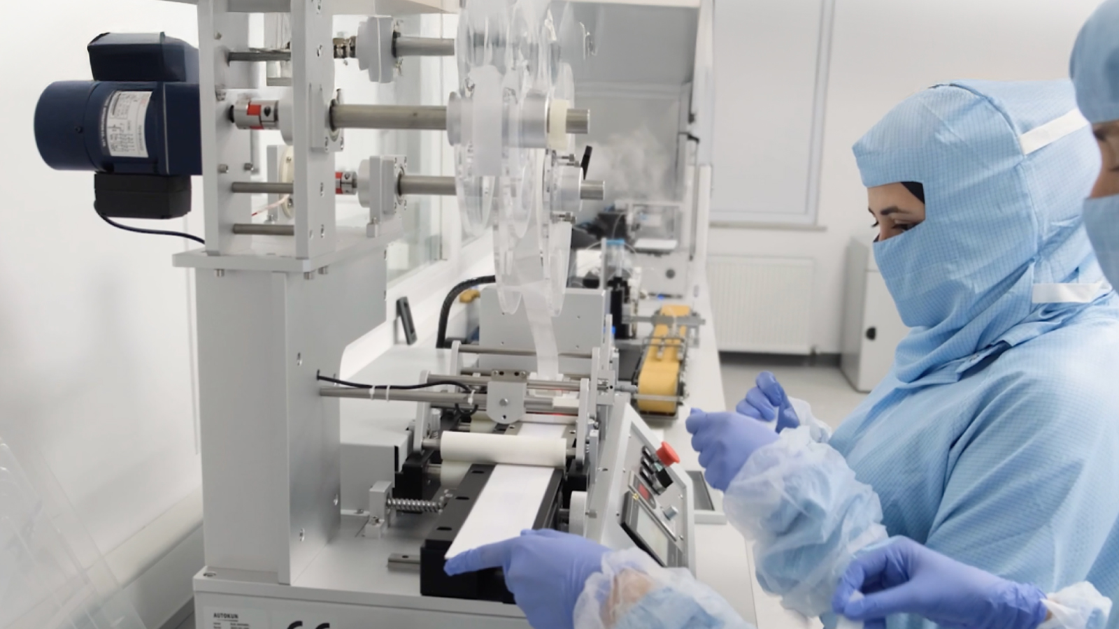
Immunoassays are widely used in diagnostic testing to detect specific biological targets through antigen-antibody interactions. Two important approaches are the lateral flow assay (LFA) and fluorescence immunoassay (FIA). While both technologies can use membrane-based test formats and specific binding reactions, they differ mainly in how the resulting signal is generated and detected.
LFA commonly produces a directly visible signal, whereas FIA uses a fluorescent label and an analyzer to measure the emitted fluorescence.
What Is a Lateral Flow Assay (LFA)?
A lateral flow assay (LFA) is a membrane-based immunoassay designed to detect a target analyte in a sample, often within minutes. The sample moves laterally across the test strip by capillary action, allowing the assay to operate without pumps or other equipment to drive sample flow.
A typical LFA strip contains a sample pad, conjugate pad, nitrocellulose membrane, test line, control line and absorbent pad. Each component has a specific function in moving the sample, facilitating the immunological reaction and producing an interpretable test result.
This simple architecture is one reason lateral flow technology is widely used in rapid diagnostic testing.
How Does a Lateral Flow Test Work?
The basic principle of an LFA can be explained in a few steps. First, the sample is applied to the sample pad and begins moving through the strip by capillary action. It then reaches the conjugate pad, which contains labeled detection components.
If the target analyte is present, it interacts with the corresponding recognition molecules and the resulting complex continues migrating along the nitrocellulose membrane. At the test line, immobilized capture molecules can retain the target complex and generate a detectable signal.
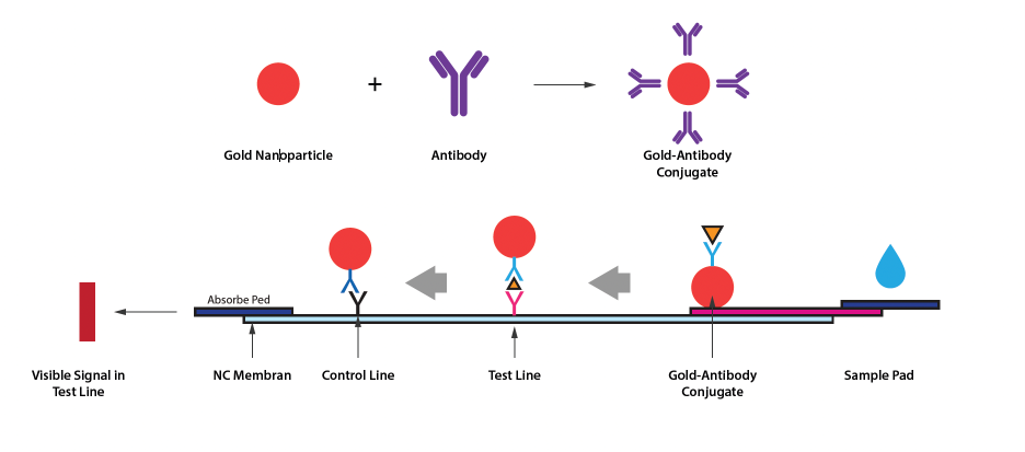
In colloidal gold-based LFAs, gold nanoparticles act as visible signal labels. Their accumulation at the test line produces the characteristic colored line associated with the assay response.
The control line provides a procedural control for appropriate test migration. Interpretation of test and control lines must always follow the Instructions for Use of the specific assay.
What Is a Fluorescence Immunoassay (FIA)?
A fluorescence immunoassay (FIA) is an immunoassay that combines specific antigen-antibody interactions with fluorescence-based signal detection. Instead of relying only on a colored line that is visually interpreted, FIA uses fluorescent labels whose emitted signal is measured by a compatible analyzer.
After the prepared sample is applied to the test device, the target analyte interacts with fluorescently labeled detection components. The resulting immune complexes are captured in the designated detection area. An analyzer then excites the fluorescent label and measures the emitted light.
Depending on the individual assay and its intended use, the measured fluorescence can be processed to provide qualitative or quantitative information.
How Does FIA Technology Work?
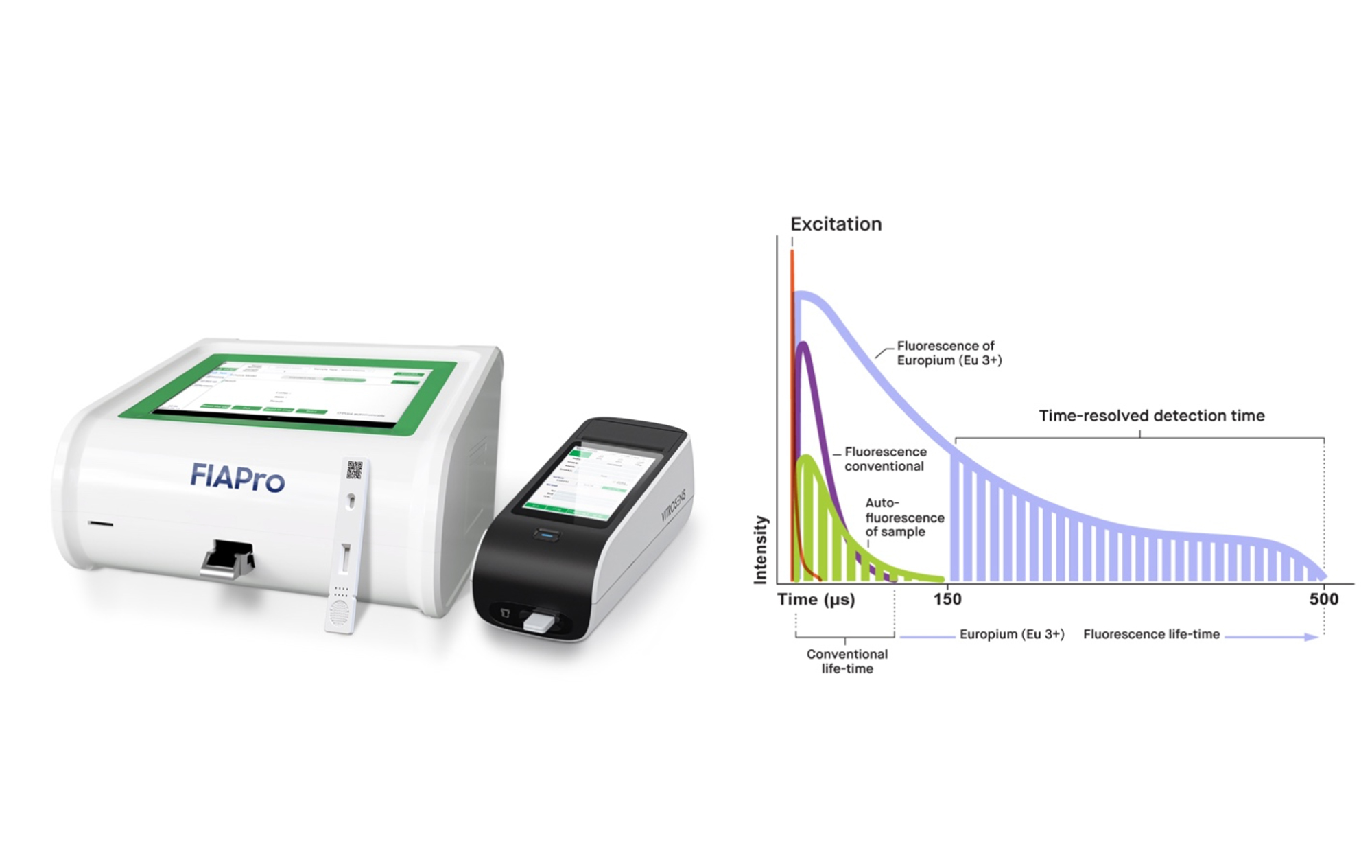
The core principle of FIA is converting an antigen-antibody reaction into a measurable fluorescent signal. After the sample migrates through the test membrane and target molecules bind to labeled detection components, the analyzer excites the fluorescent label and detects the emitted light. The measured signal is then processed according to the parameters established for the specific assay.
Some FIA platforms use time-resolved fluorescence (TRF) to further distinguish the analytical signal from short-lived background fluorescence. For example, europium chelates can provide a relatively long fluorescence lifetime, allowing background autofluorescence to decay before the relevant signal is measured.
What Is the Difference Between LFA and FIA?
The main difference between LFA and FIA is the method used to detect the assay signal. A conventional colloidal gold LFA generally produces a colored line that can be visually interpreted, while FIA generates a fluorescent signal that requires an optical reader or analyzer for measurement.
Feature | Lateral Flow Assay (LFA) | Fluorescence Immunoassay (FIA) |
Core principle | Antigen-antibody interaction | Antigen-antibody interaction |
Sample movement | Capillary action | Commonly capillary action in cassette-based FIA |
Signal label | Often colloidal gold nanoparticles | Fluorescent labels |
Signal detection | Usually visual | Analyzer-based |
Instrument required | Often no | Yes |
Result format | Commonly qualitative | Qualitative or quantitative, assay-dependent |
Typical setting | Rapid testing, laboratory or POCT depending on assay | Laboratory and POCT depending on assay |
Table 1. Comparison of lateral flow assay (LFA) and fluorescence immunoassay (FIA) technologies, including signal generation, detection method, instrumentation requirements and typical result formats.
Neither technology is universally “better” than the other. The appropriate method depends on the target analyte, required analytical performance, clinical application, testing environment and intended use of the individual assay.
Conclusion
Lateral flow assays and fluorescence immunoassays both use specific biological recognition to detect defined analytes, but they differ primarily in signal generation and measurement. LFA commonly uses visually detectable labels such as colloidal gold, while FIA uses fluorescent labels measured by an analyzer.
These differences influence how each technology is integrated into diagnostic workflows. Regardless of the technology used, results should always be generated and interpreted according to the intended use and Instructions for Use of the individual assay. To learn more about the assay and other rapid testing solutions, explore the Vitrosens portfolio or contact sales@vitrosens.com.
References
- Koczula KM, Gallotta A. Lateral flow assays. Essays in Biochemistry. 2016;60(1):111-120.
- Sajid M, Kawde AN, Daud M. Designs, formats and applications of lateral flow assay: A literature review. Journal of Saudi Chemical Society. 2015;19(6):689-705.
- Hemmilä I, Laitala V. Progress in lanthanides as luminescent probes. Journal of Fluorescence. 2005;15:529-542.


